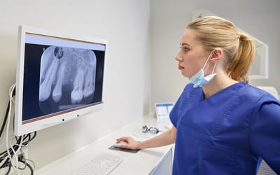
A tooth x-ray is a very safe and important tool used to diagnose dental problems. A digital x-ray scanner processes the x-ray films which requires a minimal and safe amount of radiation. Dentists can diagnose and better plan your treatment using the x-ray findings.
Tooth x-ray
Dental x-rays are the most valuable tool to diagnose:
- Cavities – X-rays will show decay in the tooth and how far the hole has extended.
- Gum disease and bone loss – If you have gum disease, the tooth x-ray film will show the extent of the bone loss. We can decide on the treatment plan according to your gum condition.
- Tooth infection – A tooth x-ray will show whether the carious lesion has gone to the tooth nerve or pulp and the extent of infection underneath the roots.
- The shape of the tooth and roots– If you need to get your tooth pulled out, the radiograph film will show the form of the tooth root. This includes whether the roots are curved, hooked or bulged.
- Lesion in the bone – infection, bone lesions or pathology will show up on the radiographs.
- Tooth fracture, root fracture or bone fracture under the gum – In most cases, we can see tooth fractures in the mouth, but we can’t see the fracture underneath the gum unless we take an x-ray.
- Teeth and root relationship to the adjacent structures– In the upper jaw, some teeth roots are closer to the maxillary sinus. In the lower jaw, the nerve is closer to the tip of the molar root.
- Missing teeth or position of the tooth– Some people have missing teeth. We can find out whether the tooth is completely missing or lying in the bone in a different angle or position. Most wisdom teeth will be impacted in the mouth. The dentist can see the position and impaction using radiographs.
- Root canal length – When performing root canal treatment, we need to determine the length of the root canal. We place a small instrument inside the nerve canal of the tooth and take x-rays to find out the correct length of the tooth’s root canal.
Types of dental radiographs
There are many types of x-ray images used to diagnose the lesion or see the structures underneath the gum. The types are:
Bitewings radiograph – The process involves small digital sensor plates or films. We place the films inside the mouth and take x-ray images. The image will show both the upper and lower teeth crowns and some parts of the gum. We use this full image to see cavities in between teeth. These images will also show us if there is any bone loss underneath the gum.
Periapical radiographs – We place digital sensors in the mouth and take pictures. This will show the full tooth, the crown, the root and surrounding bone. These pictures will show us any infection of the tooth, shape of the root and any bone pathology.
Occlusal radiograph– These sensors are slightly larger than the bitewing and the periapical sensors mentioned above. We place the sensor between the upper and lower teeth and take images. These pictures will show missing or impacted teeth as well as any bone lesion.
Panoramic, also known as OPG – The radiographic image will show the whole jaw and all the teeth. The image will show the dentist the impacted and missing teeth and any bone abnormality.
CT scan and cone beam– We take these 3D pictures before placing implants. 3D pictures help us see a particular area or problem better.
As with all procedures, we will seek permission before taking any x-rays of your mouth. If you don’t wish for us to take x-rays, you can always decline. Please note, that without taking radiographic images our dentists may not be able to detect or treat all teeth and mouth problems that you may have
Are dental X-Rays safe?
The exposure to radiation through dental x-rays is very minimal and extremely safe. If you stand in the sun for a few hours, you will get more radiation from the sun than one small dental x-ray image.
We need a very minute amount of radiation to take digital dental x-rays.
Pregnancy and dental X-rays
Dentists at Bendigo dental use digital x-ray films. This means there will be very minimal exposure to radiation. Usually, we don’t carry out routine dental radiographs if you are pregnant. If you are pregnant and have a dental emergency and it is absolutely necessary to take radiographic pictures to find out the problem, we place a lead apron on you.
This will minimise the radiation to the baby and reduce the number of exposures. If you stand in the sun for a couple of hours, you will be exposed to more radiation when compared to one digital x-ray.
